Microscope
InquiryThrough our global network of testing experts and analytical equipment including chromatography (HPLC, GC, GC/MS) and atomic absorption spectroscopy (AAS, GFA, FIAS), Our goal is to provide test services as efficiently as possible to maximize our customers' profits. For more information about our services, contact one of our experts today.
Note: this service is for Research Use Only and Not intended for clinical use.
Optical microscopies have been extensively used in microbiology, biotechnology, pharmaceutical research, and food testing. The advanced microscopy techniques nowadays allow observations of organelles or biomolecules in living cells, which greatly facilitates the study of various biological processes.
Super-resolution Multiphoton Confocal Microscope
Leica TCS SP8 STED 3X allows the study of sub-cellular structures and cell dynamics at the nanoscale. The system is capable of continuous-wave Stimulated Emission Depletion (cwSTED) and gated STED imaging, which improves resolution and live cell imaging capabilities. System also can be applied to standard confocal imaging for structures study.

Vega LaB6-SEM
VEGA LaB6 scanning electron microscope has both high and low vacuum modes. It is suitable for the observation of surface morphology of conductive and non-conductive materials. With enhanced depth of field display mode, depth of field up to 7mm or above, dedicated to the observation of high and low drop samples, such as metal fracture, failure analysis, the secondary electron image and backscattered electron image of surface morphology can be obtained.

Scanning Transmission Electron Microscope (S/TEM)
The Talos F200X G2 is a 200 kV FEG Scanning Transmission Electron Microscope (S/TEM), which is designed for fast, precise and quantitative characterization of nano-materials.

Field Emission Scanning Electron Microscope (JEOL JSM-7800F Prime)
SEM is the main instrument for microstructure analysis, which has been widely used in metallurgy, minerals, biology and other fields. JSM-7800FPRIME delivers the world's best resolution with the incorporation of the newly-developed, super-high resolution Gentle Beam (GBSH). In addition, the maximum probe current of the In-lens Schottky Plus gun has been increased from 200 nA to 500 nA.

Atomic Force Microscope (AFM)
AFM is a surface measurement technique that is based on the interaction of a tip with the surface of the sample. This technique allows the surface analysis of samples with nanometric or even atomic resolution. It has the possibility of making measurements without any previous treatment of the sample to be measured, and without the need to use vacuum. Multimode AFM Nanoscope III A (Bruker) is equipped with three 1 μ, 15 μ and 150 μ scanners.

Field-emission Scanning Electron Microscope (FE-SEM, *NOVA NanoSEM 230)
Nova NanoSEM 230 scanning electron microscopy coupled with large area SDD spectrometer and rapid electron backscatter diffraction (EBSD) can be used for morphology observation, element composition and distribution analysis, structure and orientation analysis, such as metals, nanoparticles and powders, nanotubes and nanowires, porous materials (such as silicon, hydroxyapatite), plastic electronic devices, glass matrix materials, organic materials, macromolecule materials, diamond, stone films, semiconductor cross-section, crystal materials, biological samples, all kinds of film materials and cross sections.

MIRA3 (SEM)
MIRA3 is a high performance SEM system which features a high brightness Schottky emitter for achieving high resolution and low-noise imaging. MIRA3 offers all the advantages that come with the latest technologies and developments in SEM; delivering faster image acquisition, an ultra-fast scanning system, dynamic and static compensation and built-in scripting for user-defined applications.

Sirion 200 Field Emission Gun Scanning Electron Microscope (FEGSEM)
The FEGSEM facility is based on a FEI Sirion 200 which is a is an ultra-high resolution Schottky field emission scanning electron microscope and is ideal for studying materials on the nanometre scale. The microscope is equipped with energy dispersive X-ray analysis (EDX) and electron backscattered diffraction (EBSD).

Leica DM 4000
Optical microscope is the most basic means of morphology characterization, which can be used to analyze the morphology and structure of materials in a lower multiple and a larger range, and to observe some contrast which is not easy to be observed by SEM. It is a good supplement to SEM system. DM4000M can be used to identify and analyze the organization of various materials as well as the quality inspection and control of a series of production processes in inspection, casting, pressure processing, and heat treatment of raw materials. It can also be applied to the development and use of new materials and new technologies.

Environment Control Scanning Probe Microscope
The scanning probe microscope (Nanonavi E-Sweep) can be used to measure the surface nanostructures of samples under variable temperature and vacuum conditions, which provides an important condition for the study of the structural changes at different temperatures and atmospheres.

Raman Microscopes
The SENTERRA is a high performance Raman microscope spectrometer designed for demanding analytical applications in both the R&D and routine laboratory. SENTERRA integrates a multi laser Raman spectrometer onto the confocal optical microscope. The spectrometer part is integrated in between the base and the binocular of the microscope. Due to this compact design, the beam path is kept very short, which accounts for the high stability and sensitivity of the system.

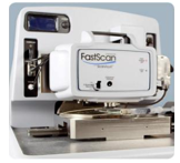
Dimension FastScan Bio™ Atomic Force Microscope (AFM)
The Dimension FastScan Bio™ Atomic Force Microscope (AFM) enables high-resolution research of biological dynamics, with temporal resolution of up to 3 frames-per-second for live sample observations. What's more, it does this while making the AFM easier to use than ever before.
Biology Transmission Electron Microscope
The FEI company's Tecnai G2 spirit Biotwin biotype TEM adopts their patented technology, Biotwin, which greatly improves the contrast of the TEM. The biological transmission electron microscope is suitable for observing the ultrastructures of biological samples such as bacteria, viruses, tissues, cells and organs, as well as organic samples such as polymers and micelles.

Olympus BX61
Olympus BX61 motorized upright microscope with fluorescence and phase contrast optics for immunofluorescence imaging. The BX6 incorporates infinity-corrected optics, motorized Z-axis (focus), and equipped for brightfield, Nomarski DIC, and widefield epi-fluorescence techniques.

Services
Our services have the following advantages:
Tailored experimental design
Applicability for a wide variety of sample types
One-stop service from sample preparation to data acquisition
Short detection cycle
Competitive price
Years of experience
Do not know how to place an order, please refer to the flow chart shown below.
Submit quotation request |
A technical manager will contact you within 24 hours |
You will review and approve the final price and place an order |
Confirm with you and make the payment |
Instruct you to ship your samples and form |
Analytic report delivery |
